The
central theme of Dr. Hsiai's research program is primarily
on hemodynamics and mechanobiology via the application of engineering
principles and techniques, including Bio-MEMS and nanotechnology to
study oxidative stress and to link physical and chemical properties
with vascular biology. Using
a micro-fluidic flow model, Dr. Hsiai's lab is investigating the
mechanisms whereby hemodynamics regulates the development of coronary
artery disease. His group is linking the effects of real-time shear
stress on cardiac cell dynamics using intravascular polymer sensors,
molecular biology techniques, and proteomics. Using zebrafish
heart model, the group is developing micro-ECG for longitudinal
monitoring for cardiac arrhythmia. The current research objective is to
study vascular inflammatory responses in the context of electrochemical
properties and vascular calcification.
Our Projects
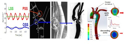
Hemodynamics
and Cardiac Development
Electrical
and Mechanical coupling of Zebrafish Hearts
Hemodynamic
forces
are intimately linked with cardiac
development and normal function. Circulating blood imparts fluid shear
stress on the walls of the heart during cardiac contractions. Our group
is interested in investigating the link between fluid shear stress and
cardiac development in a Zebrafish model. In collaboration with
Professor Chih-ming Ho, we are acquiring real-time 3D beating heart
images using selective plane illumination microscopy (SPIM). This
imaging method allows us to determine boundary conditions from image
data to prescribe wall motion for computed fluid dynamic (CFD)
simulations. In collaboration with Professor Alison Marsden from UCSD,
we are assessing hemodynamics and cardiac morphogenesis via the use of
moving boundary Computational Hemodynamics (CFD).
Our laboratory has
been inspired by the developmental
biologists for their seminal contributions to cardiovascular medicine
using the zebrafish heart model. We and others have developed micro-ECG
strategy (Dr. Tai at Caltech) to assess conduction phenotypes in adult
and embryonic zebrafish. In collaboration with the zebrafish heart
experts, we observed changes in conduction phenotypes in the
regenerating myocardium in response to ventricular injury. In
collaboration with Dr. K. Kirk Shung, NIH Director of Ultrasonic
Transducers Resource Center, we are demonstrating mechanical phenotypes
in terms of ventricular compliance (E- and A- waves) with high spatial
and temporal resolution. In collaboration with zebrafish experts, Dr.
Ellen Lien at Los Angeles Children's Hospital, Dr. Jau Chen at UCLA,
Dr. Xiaolei Xu at Mayo Clinic, and Dr. Neil C. Chi at UCSD, we are
assessing the biomechanical mechanisms underlying the
functional/physiological phenotypes in the regenerating myocardium by
incorporating microelectrode arrays and high frequency ultrasonic
transducers, histology and optical voltage mapping conduction. These
fundamental findings will pave the way for early detection, monitoring,
and management of aberrant electrical signals in neonatal models of
tissue regeneration.
Vascualr
Repair Related in Shear Stress and Oxidative Stress
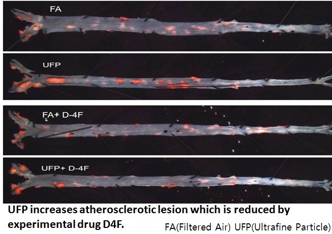
Urban
Air Pollutants Ultrafine Particles and Vascular Biology
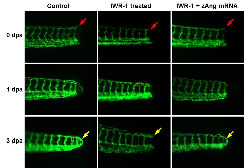
Wnt-Ang-2
signaling and vascular endothelial repair.
The tails of transgenic Tg (kdrl:GFP) zebrafish embryos were amputated
at 72 hpf . At 0 day post amputation (dpa), the red arrow pointed to
the site of injury. At 1 dpa, initiation of endothelial repairs was
present. At 3 dpa, complete tail repair was observed, as indicated by
the yellow arrow.
Chronic exposure to
ambient urban ultrafine particles (UFP, dp < 150 nm) is an
emerging environmental risk factor associated with increased morbidity
and mortality. This issue is particular relevant in large cities that
rely on automotive transportation such as Los Angeles. In a
collaborative effort with Professors Constantinos Sioutas and Professor
Celeb Finch from USC and Professors Mohamad Navab, Jesus Araujo, Linda
Demer, and Prof. Yin Tintut from the UCLA School of Medicine, we are
assessing the molecular mechanisms underlying multi-organ system
effects in response to ambient air pollutant exposure. Our
collaborations have culminated in novel signal pathways leading to
endothelial cell dysfunction and CVC cell calcification, as well as
lipid peroxidation and reduced HDL anti-oxidant capacity in the
LDLR-null mouse model.
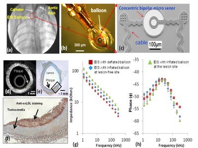
Integrated
intravascular sensors to assess unstable plaque
Hemodynamics
& Mechanobiology of Endothelium
Our group have
developed the first quantitative micro-technological approach to
measure in real-time changes in intravascular shear stress in the New
Zealand White rabbits and swine models. We have further applied
microelectromechanical systems (MEMS) and nano-scale sensors for
real-time quantification of shear stress and oxidative stress with
pathological significance to the initiation of inflammatory resposnes.
Our micro-enabled technology (e.g., MEMS shears stress sensors and
concentric bipolar microelectrodes), molecular tools (e.g.,
adenoviruses to over-express Mn-SOD, small interference RNAs, flow
cytometry to measure mitochondrial redox status), as well as transgenic
and knockout animals
Our group has
demonstrated the first quantitative
approach to changes in intravascular shear stress (ISS) with
vascularnulloxidative stress in New Zealand White (NZW) rabbit and
swine models. We have demonstrated that spatial (/x) and temporal (/t)
variations in shear stress modulate post-translational oxidative
modifications of low density lipoprotein protein (LDL) and
mechano-signal transduction of mitochondrial redox states.
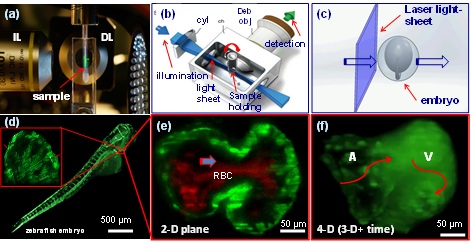
Fluorescent
light-sheet microscopy to image 3-D zebrafish embryo and beating heart
Ultramicroscopy
with Super-Resolution and Clearing Techenique
(a) The sample is placed at the intersection of
illumination lens (IL) and detection lens (IL) in our LSM system. (b)
LSM applies a laser light-sheet to illuminate the sample. The
illuminated planes are orthogonally detected by the detection
objective. Cyl: cylindrical lens. Deb obj: detection objective. (c) A
schematic diagram illustrates a sheet of laser light transverses the
embryo. (d) The entire embryo can be imaged at the micron scale within
30 seconds. Inset reveals the trabeculated endocardium. (e)
Magnification of the heart reveals the fli-1-eGFP-labeled endocardium
and DsRed-labeled red blood cells throughout the cardiac cycle. (f) 4-D
LSM image reveals a beating heart.
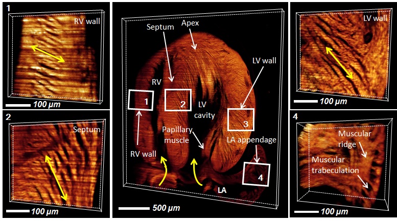
By slicing and applying the super-resolution to increase the
image resolution for optically cleared large-scale neonatal mouse
hearts (several milimeter scale),
followed by volumetric rendering, we demonstrated the use of 4x
objective with a numerical aperture of 0.13 to resolve the single cell
level of cardiac trabeculation and changes in orientation of myocardial
fibers from left ventricle to septum, to right hearts.
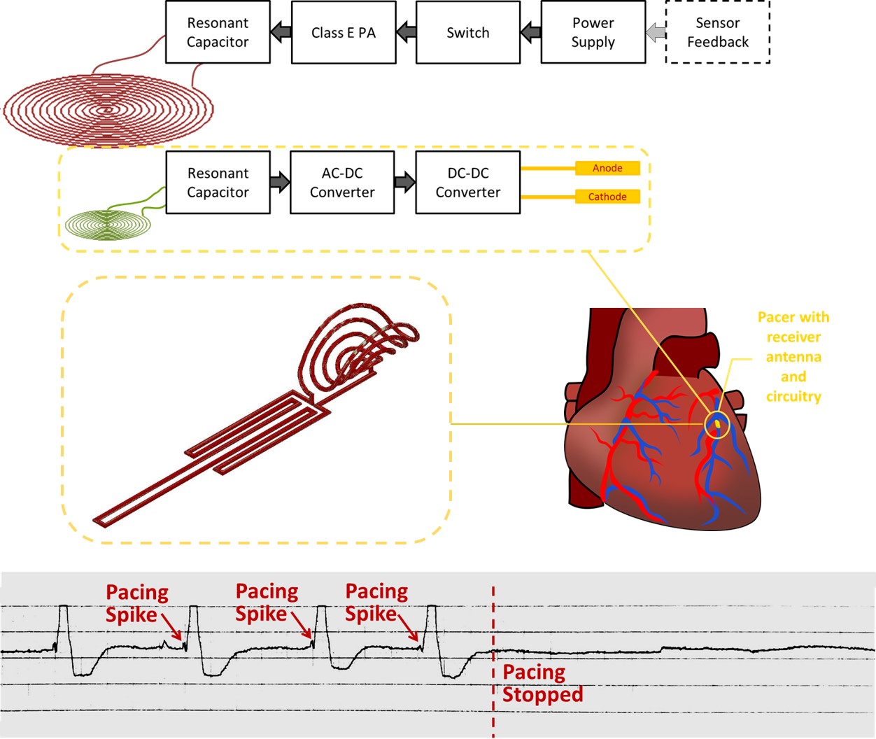
Despite great advancements in implantable cardiac pacemaker
technology, complications from pacemaker leads continue to compromise
nearly 10% of all implants. This has motivated significant research for
the development of leadless devices. These include battery-based
systems, power harvesting devices, and stimulators wirelessly powered
through radiofrequency radiation. Our research is focused on the
development of an inductive power transfer system with a remote
stimulation control system. Our proposed system is capable of
significantly improving power efficiency and reducing tissue energy
absorption in a highly asymmetrical system in which the pacer is small
enough to be intravascularly deployed and implanted with a stent-like
fixation mechanism.